• Hello guest! Are you a Tegu enthusiast? If so, we invite you to join our community! Our site is specifically designed for you and it's a great place for Tegu enthusiasts to meet online. Once you join you'll be able to post messages, upload pictures of your Tegu and enclosure and have a great time with other Tegu fans. Sign up today! If you have any questions, problems, or other concerns email [email protected]!

MBD, prolapse, and feeding

Guman

Active Member
Messages
204
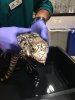
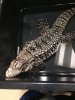
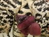
Ok, looking for some help. Last wed. my boy prolapsed and we cleaned him up and was able to reduce at home. Then, on Sunday night late he reprolapsed. Was worried about an are that looked purple like maybe dead bowel. So, we loaded him up drove 5.5 hr. In a snow storm to take him to Colorado State University. He was seen by a general vet who consulted exotic vet. The exotic team stated it was just congested and to use sugar to reduce and transfer to exotic team in AM. She could not reduce, so he waited till exotics came in. Latter, he was sedated and reduce with two stiches placed in vent. We did xrays, blood work, fecal sample, fluid, antibiotics, and pain meds. My baby is still in hospital. The only parhology found was low white blood cell and the said bone density looks bad. (MBD)?

Ok so sorry this is long a few questions.

I am lost on the MBD. He has UVB changed every 6 months. He is fed raw meat and will not eat fruit or veggie. We use multi vit. Supp. Blood did not show Ca+ or phosphorus abnormalities on blood.

Temps high 110 low 75-80. Humidity 40-90%

But, he hides most of the time so ok maybe not enough light.

They only think MBD bc of xray. But I question if KV'S on xray was set too high? He is not my first large lizard. Have a fat sassy healthy Iguana girl.

They told me he has not reprolapsed since a small one after the stiches was put in on Monday night. He has had a small bm yesterday with no problem. ( still has some frank gi bleeding, they think bc of ulcers on colon). Fecal shows no worms or high bactirial count. They want to do an Ultra Sound for $350. I have a bill of $1500 already. The vet said no raw diet and fed wet cat food in syg. Yesterday.

Looking for some gu owners ideas. I have some Oxbow carnivore care coming overnight. I am thinking about holding off on ultra sound.

I want the best. Feeling like a failure! I have tried so hard to care for him. Was able to stop the MBD that my iguana had when I purchased her. She laid 18 healthy strong eggs last week. UVB for him is a zoomed reptisun no diffuser on at 12 in away. If this is MBD all I can come up with is removing his hide.

So, what do I feed when he comes home?
Would you wait on Ultra Sound?
Ideas to get him more UVB?
Can low WBC be a sign of infection in gu's?
Anyone have a MBD baby how have you helped improve their life?
He will be on antibiotic injections and pain inj. Any advice?

Thank you!! For any advice!
 

beardeddragon111

Active Member
Messages
371
Is your UVB the tube kind or coil kind. The coil UVB don't work well. IMO MVB's are best. As far as MBD goes, if you have the wrong UVB, and your feeding only raw meat, there's your problem. You'll want to switch to whole prey, mice, rats, chicks, etc. I couldn't help you on the white blood cells and prolapse's. Sorry about that. Hope your tegu does well :).
 

Guman

Active Member
Messages
204
Light is a T8 tube, zoomed 18", no reflector, plastics off.

Yes, he was not fed whole prey. I am concerned about feeding pinkies ect. With the prolapse. Would ground whole pray be ok? I did order some Oxbow carnivore care for when he gets home today. Wanted to have that on hand incase he needed syg. Fed.

The vet has been doing wet cat food.

I also will be getting a difrent humidity monitor! I have an infrared temp gun.
 

beardeddragon111

Active Member
Messages
371
Whole ground meat is fine. It's confusing to me then that he'd have MBD. Where did you get the lizard? Is it possible you purchased him sick? Sounds like he's very young if he can't eat pinkies. Otherwise the care sounds ok.
 

Guman

Active Member
Messages
204
I have had him since he was a green head hatchling. He was a pet store save. They was going to kill him bc they said he was mean. Cant wait to get him home. Here is a pic of my boy at vet.
 

Attachments

  • IMG951743.jpg
    IMG951743.jpg
    200.1 KB · Views: 110

Guman

Active Member
Messages
204
Whole ground meat is fine. It's confusing to me then that he'd have MBD. Where did you get the lizard? Is it possible you purchased him sick? Sounds like he's very young if he can't eat pinkies. Otherwise the care sounds ok.

Not that he is really young. Just thinking whole bone may be hard on system after having rectum prolapsed.
 

Attachments

  • 20180121_225519.jpg
    20180121_225519.jpg
    3 MB · Views: 60

beardeddragon111

Active Member
Messages
371
Not that he is really young. Just thinking whole bone may be hard on system after having rectum prolapsed.
Good point. If you feel the need try liquid calcium. But that shouldn't be necessary with whole ground meat

Sent from my SAMSUNG-SM-G920A using Tapatalk
 

Gary

Member
Messages
83
It won't be bone when it hits the lower bowels, but listen to the vet recommendation on wet cat food for now. Whole prey has a lot of benefits. Work fruits and veggies on and around the meal in small portions. Hopefully he'll get a taste for the variety. He may just be very conditioned to ground meat. My little guy would only eat ground meat because of his breader, but I was able to slowly transition him over 3 months. Once your gu has recovered, skip a day of food to drive appetite up then offer something new.
 

Zyn

Well-Known Member
Messages
609
I'd also try some live minnows and crayfish. My little guy loves him some minnows. As far as MBD is his calcium low? That's what the whole prey is for the bones hairs nails everything is calcium. I own 5 aches 3 of which are woodland with a large steam this last summer he was eating all kinds of wild caught crayfish, mennows, tadpools. Also have a small Breeder colony of dubia and rats feeding off most babies. He's to the point where be only wants whole prey. The small rats are pre-killed.

My only guess is he's having issues absorbing calcium. He looks healthy in the pictures doesn't look dehydrated, whole prey also offers all the minerals needed for proper calcium absorption.

Try lowering the UVB sourcs 10-8 inches but that's just a guess.

Also beef liver will do wonders.

Edit: at his size he would be able to put down small rats and chicks for sure. The chick are
Full of calcium with the beaks and feathers and what not.
 

Guman

Active Member
Messages
204
It won't be bone when it hits the lower bowels, but listen to the vet recommendation on wet cat food for now. Whole prey has a lot of benefits. Work fruits and veggies on and around the meal in small portions. Hopefully he'll get a taste for the variety. He may just be very conditioned to ground meat. My little guy would only eat ground meat because of his breader, but I was able to slowly transition him over 3 months. Once your gu has recovered, skip a day of food to drive appetite up then offer something new.
He has been eating bonless chicken, beef, pork, salmon, tilapia, shrimp, and ect.

We have made our 5.5 hour trip to CSU to pick him up from the exotics team. Just waiting on them. They had clinicals at the Colorado Springs Zoo today. During our trip we have talked about maybe changing him to a mecury vapor bulb and just getting our own meat grinder for him. This way he can have the benefits of whole pray while his gut heals.
I have to say this vet would have to show me research proving cooked food is better than raw. Right now I belive in raw.
 

beardeddragon111

Active Member
Messages
371
He has been eating bonless chicken, beef, pork, salmon, tilapia, shrimp, and ect.

We have made our 5.5 hour trip to CSU to pick him up from the exotics team. Just waiting on them. They had clinicals at the Colorado Springs Zoo today. During our trip we have talked about maybe changing him to a mecury vapor bulb and just getting our own meat grinder for him. This way he can have the benefits of whole pray while his gut heals.
I have to say this vet would have to show me research proving cooked food is better than raw. Right now I belive in raw.
Cooked food is definitely not as good as raw. If you don't want to get a meat grinder, check out hare-today.

Sent from my SAMSUNG-SM-G920A using Tapatalk
 

Guman

Active Member
Messages
204
Gave him his shots this morning, soaked, and fed. I do not think he will take these shots well by the end.
 

Guman

Active Member
Messages
204
At least you'll know he's doing better if he starts trying to bite your fingers off.

Sent from my SAMSUNG-SM-G920A using Tapatalk
True that! He was good about the syringe feed today. After he found out it eas food he would open his mouth bite the syringe and let me push the food in.
 

dpjm

Active Member
5 Year Member
Messages
378
Guman, I want to congratulate you on you commitment. Long drives and large vet bills... many would not go to that trouble.

I also wanted to explain how the x-ray could show evidence of MBD but the blood work shows normal calcium levels. Blood is the most important place for calcium to be in the body and so blood calcium is tightly regulated by the endocrine system, i.e., hormones. If blood calcium becomes low because it is not being supplied in proper amounts from the diet (or for other reasons) then the body will produce hormones that cause release of calcium from bones into the blood. Think of the bones not only as structural components to the body but also as calcium storage banks, where the body can make withdrawals and deposits as needed. There are essentially endless funds available here.

So when diagnosing MBD, xrays are a much better tool than blood tests that check calcium levels.
 

Guman

Active Member
Messages
204
Dpjm thank you for that explanation. One of my questions to the vet was, why we were not checking for hyperparathyroidism? As you pointed out hormones are released to stimulate the release if ca+ from bones. In addition, during my research it sounded like hyperparathyroidism would be present if the kidneys have been effected by the MBD. Does anyone here know if I am correct?


Renal Disease in Green iguanas and other Reptiles (Kidney disorder)
Book: Reptile Medicine and Surgery. Douglas R. Mader, MS, DVM, DABVP Diplomate, American Board of Veterinary Practitioners (CA) Fellow, Royal Society of Medicine Marathon Veterinary Hospital Marathon, Flori.da SAUNDERS. With 72 Contributing Authors

Continued strides in captive husbandry and nutrition of reptiles have resulted in many animals surviving to adult and geriatric ages. Nevertheless, renal disease remains a major cause of morbidity and mortality,particularly among green iguanas, and continues to be of significant concern to veterinarians and reptile keepers alike. Inadequate captive husbandry, poor nutrition, and secondary nutritional hyperparathyroidism are considered to becoming predisposing factors to chronic renal disease, and acute renal failure tends to be the result of infectious or toxic causes and more sporadic in nature. Although most of the information presented here relates to lizards, and especially the Green Iguana (Iguana Iguana), much has direct relevance to reptiles in general.

Renal Disease (explanations added my me)
A variety of renal disease have been described in iguanas and other reptiles, including:

  • Renal cysts - kidney cysts
  • Interstitial nephritis - kidney disorder in which the spaces between the kidney tubules become swollen (inflamed)
  • Glomerulonephritis - a type of kidney disease in which the part of your kidneys that helps filter waste and fluids from the blood is damaged.
  • Pyelonephritis - kidney infection
  • Glomerulosclerosis - hardening of the glomerulus in the kidney. It is a general term to describe scarring of the kidneys' tiny blood vessels.
  • Nephrosclerosis - Hardening of the walls of the small arteries and arterioles
  • Glomerulonephrosis - non-inflammatory disease of the kidney
  • Tubulonephrosis - lack of oxygen to the kidney tissues
  • Renal edema - swelling caused by fluid retention
  • Amyloidosis - amyloid proteins are abnormally deposited in organs or tissues
  • Gout - uric acid builds up in blood
  • Bacterial nephritis - localized bacterial infection of the kidney presenting as an inflammatory mass
  • Tubular adenoma - a type of colon polyp that may turn cancerous
  • Adenocarcinoma - a cancer of an epithelium that originates in glandular tissue
  • Metastatic adrenal (intrarenal) carcinoma - cancer


In the terminal stages,aberrant calcium metabolism may cause metastatic calcification of the cardiorespiratory and gastrointestinal system (Figure 66-2, B). High-protein diets are considered a predisposing cause of hyperuricemia, gout, and nephrosis in herbivorous reptiles,and modern texts now recommend avoidance of such foods.

Blockage of the renal tubules with secreted urate is another process that impair renal function and has been associated with chronic dehydration in snakes. The maintenance of the high-humidity, rain forest species in dry captive conditions is suggested as a cause of chronic dehydration and nephropathy(kidney disease).

CLINICAL INVESTIGATION


Currently, diagnoses of renal disease in reptiles is made on the basis of history, physical examination, haematology, biochemistry, urinalysis, diagnostic imaging, and ultimately renal histopathology and micro biology. However, the recent advent of measurement of iohexol excretion to determine GFR offers the thirst practical means for assessment of renal function.

Anamneses (patient case history)

A detailed history is most important in investigation of any disease; moreover, in cases of suspected renal disease history can often differentiate between true acute renal disease and chronic renal failure. Usually little history is associated with acute renal disease. In most cases, the lizard present with acute onset depression, anorexia, and usually cessation of urine and urate output. Such cases often involve individuals that are poorly managed in unhygienic conditions. Recent exposure to nephrotoxins including aminoglycoside antibiotics and high doses of vitamin D3 may be inferred from a detailed case history and previous medical or owner records. Total water deprivation, severe dehydration, or haemorrhage could also theoretically lead to poor renal perfusion and acute renal failure.

Reptiles with chronic renal disease often have had long term mismanagement. High protein diets rich in purines and in particular the use of canned dog or cat food an lead to abnormal hyperuricemia in herbivores, increased demands for effective urate excretion. Likewise, persistently inadequate humidity or inappropriate water provision (e.g., water bowl instead of daily spraying) can result in chronic dehydration. The regular use of oral vitamin D3, as a substitute for broad -spectrum lighting can cause nephrocalcinosis (too much calcium deposited in the kidneys), and reptiles that recover from secondary nutritional hyperparathyroidism may have sustained chronic renal damage from the cytotoxic effect of excess parathyroid hormone. Affected animals tend to have a more protracted history, including deteriorating body condition, capricious appetite, and lethargy that may extend over weeks or months, and as a result, most cases present dehydrated and emaciated. Only occasionally do owners report polydipsia (excessive thirst) or polyuria (excessive or abnormally large production or passage of urine).

All words in ( ) are added by me to give an explanation of the medical word.

Physical Examination

A thorough physical examination is always indicated and should include an accurate measurement of weight. Reptiles with severe renal compromise present in a depressed and weakened state (see figure 66-2). ln case of acute disease, the animal often presents in good body condition, whereas the chronic renal case is likely to be significantly underweight. Dehydration may be inferred from reductions in skin elasticity, salivary (saliva), and ocular (eye) secretions.

Pharyngeal edema (throat swelling) is not uncommon, and where digital palpation of the kidneys per cutaneous or per cloaca is possible, the size, shape, and contours of the kidneys can be determined (Figure 66-3) and should be a part of every physical examination. Pronounced renomegaly (enlargement of kidneys) may cause constipation and cloacal prolapse.


Routine Blood analysis

Blood collection and analysis is an essential part of the diagnostic investigation. Preference should be given to generating normal reference data for each individual animal when healthy (i.e yearly health examinations and blood screens).

Hematology (The study of blood)

Pathologic elevations of hematocrit (red blood cells) are usually related to dehydration. The clinician must also be aware that chronic renal disease may lead to a nonregenerative anemia (failure to produce red blood cells in the bone marrow) that may mask hemoconcentration (an increase in the concentration of blood cells). Unless immunosuppression is present, acute infection or inflammation usually results in heterophilia (antibodies) or azurophilia. In cases of chronic renal disease, a decreased, normal, or mildly increased total white blood cell count is more common, with monocytosis (increased white blood cells) not an unusual feature. Reptiles constantly exposed to temperatures below the preferred optimum temperature zone may become immunocomprimised and fail to show an appropriate leukocyte (white blood cell) response, even in the face of overwhelming infection. (When a reptile are kept at too low temperatures their immune system drop and they cannot produce white blood cells to fight a disease).

Ultrasonography


Ultrasonography, with a 7.5-mHz to 10-mHz sector transducer, permits an appreciation of normal tissue and mineralization, cysts, and other gross pathologic changes (Figure 66-1.2). Ultrasonography also aids kidney visualization for transcutaneous biopsy.


Conclusion

Renal disease remains an important disease process of captive reptiles, especially the Green Iguana. In cases of chronic renal disease, clinical signs and biochemical changes are unlikely to become apparent until late in the course of disease. Iohexol clearance, at least in the iguana, appear to be a safe and practical method for estimation of GFR (glomerular filtration rate), and for the first time, renal function can be evaluated in practice.

Endoscopic evaluation provides a safe and effective means for visualization and biopsy of kidneys, and this technique has again been validated, at least in the iguanas and chelonians. Apart from hyperparathyroidism, chronic water deprivation also appears to be a common historic factor in certain rain forest adapted species. As a folivore originating from the high humidity rainforests of Central and South America, water recovery is not considered to be an adaptive stress response in Iguana iguana, and therefore, renal anatomy and physiology are considered to be nonspecialized compared with more arid or aquatic reptiles. In addition, arboreal reptiles do not voluntarily drink from open water but instead imbibe rain or dew droplets from foliage. Maintaining such species in low relative humidity with a water bowl from which to drink is likely to both increase insensible water losses and interfere with normal water intake
 

Guman

Active Member
Messages
204
Metabolic Bone Disease - Nutritional secondary hyperparathyroidism
Book: Reptile Medicine and Surgery written by Douglas R. Mader, MS, DVM

The term metabolic bone disease (MBD) is not actually a single disease entity but rather a term used to describe a collection of medical disorders that affect the integrity and function of bones. Many different types of metabolic bone disease affect both animals and humans alike. In an effort to maintain purity in medical description, the terms metabolic bone disease and MBD should NOT be used to describe diseases of reptiles unless accompanied by a qualifier such as "nutritional" or "renal" (e.g., The green iguana had metabolic bone disease of nutritional origin). Hereafter, MBD of nutritional origin is referred to as NMBD and of renal origin as RMBD. Nutritional secondary hyperparathyroidism (NSHP) is the most common MBD diagnosed in captive herpetofauna.

Nutritional secondary hyperparathyroidism = nutritional metabolic bone disease and is a clinical manifestation of prolonged hypocalcemia. Hypocalcemia = imbalance of calcium and phosphorus in the diet and / or lack of activated vitamin D3 through UV / Sun.Hypocalcemia is when plasma calcium concentration is less than 8 mg / dl.

Low intake of calcium (directly or from vitamin D3 deficiency) stimulate parathyroid hormone (PTH) secretion and calcium is mobilized from bone to maintain normal blood calcium levels.


NUTRITIONAL SECONDARY HYPERPARATHYROIDISM - NSHP

"Rubber jaw" is the lay term often used in the pet stores and herpetologic societies. Although this term is not technically correct, the origin of "rubber jaw" comes from the fact that animals with NSHP often have grotesque facial deformities that usually affect and soften the lower jaw (Figure 61 -1).


Nutritional secondary hyperparathyroidism occurs as a result of dietary or husbandry mismanagement. The most commonly implicated factors are a prolonged deficiency of dietary calcium or vitamin D3, an imbalance of the calcium-phosphorus ratio in the diet (usually an excess of phosphorus), or inadequate exposure to ultraviolet (UVB) radiation in diurnal animals.


NSHP is common in the green iguana, and although their diets have been studied in the wild, nutritionally balanced captive diets, coupled with comprehensive applied husbandry in captivity, have yet to be fully elucidated.

Under natural conditions, the body synthesizes its own vitamin D3 when it is exposed to sunlight. Light from the ultraviolet spectrum (290 to 320 nm) reacts with the body's skin to convert cholesterol to the inactive form of vitamin D. This vitamin D is then converted to a hormone called 1,25 dihydroxycholecalciferol (l,25-DHCC) through pathways involving the liver and kidneys. The active form of 1,25-DHCC is used by the body to facilitate absorption of calcium from the intestinal tract.

Animals in a captive environment do not always get the proper exposure to natural unobstructed sunlight. An alternative to natural sunlight is artificial light that mimics the spectrum of natural light. A number of artificial
lighting systems are available that provide an ultraviolet light spectrum that simulates natural sunlight. Although they are advertised as mimicking natural sunlight, one should note that not all of these lights
actually produce the necessary wavelengths required to stimulate vitamin D synthesis in the skin. Even with the best lights on the market, one should remember that they are NOT a replacement for natural sunlight. In addition, when positioning these lights in the animal's cage, placement of the lights close enough that they are still effective is crucial.


Physiologic Response to Nutritional Secondary Hyperparathyroidism

The parathyroid hormone regulates calcium and phosphate levels and helps to maintain these levels. In NSHP, an excessive production is seen of parathyroid hormone from the parathyroid gland in response to the diet / management-induced hypocalcemia. Calcium is resorbed from the bones to compensate for the deficiency. The resulting osteopenia (bone mineral density is lower than normal) weakens the integrity of the bones. If this condition occurs in a young growing animal, it is called rickets. If it occurs in an adult, it is referred to as osteomalacia.

Laboratory findings of low normal blood calcium levels are common in NSHP. As the blood calcium levels drop,
parathyroid hormone (PTH) is released from the parathyroid gland, which increases blood calcium by increasing bone resorption, increasing renal tubular reabsorption of calcium, and in simultaneously increasing phosphate excretion in the urine. (see Figure 61 -3).


Calcium deficits cause partial depolarization of nerves and muscle because of an increase in the threshold potential. "Playing the piano" is a common phrase used to describe the rhythmic twitching of the digits seen in iguanas. An increase in neuromuscular excitability caused by hypocalcemia can account for the reptile's spastic tremors. Snakes fed a "pinkie"-only diet (consisting of only neonatal mice) have been reported to exhibit signs of tremors, disorientation, andataxia. Cloacal prolapse from hypocalcemia is a common finding in the young Iguana.


Young animals with active bone growth are the most affected. No typical presentations of the disease are found. Affected animals may show any or all of the following: deminirelized eggs (incomplete calcification, weak eggshells), and eventuality hypocalcemia (muscle tremors, seizures). Bone lesions are common in the long bones, mandible, skull, and vertebrae (Figures 18-47 through 18-49).


Thickening and swelling of the long bones and mandible (fibrous osteodystrophy), pathologic fractures of the long bones and spine (generally seen with 40% or greater loss of bone mass), horizontal (rather than the normal vertical) rotation of the scapulae -(shoulder blade), tetany (clinical hypocalcemia), muscle fasciculations (contraction), hyperreflexia, cloacal or rectal prolapse, anorexia, in ability to ambulate (carpal walking), and stunted growth. (Figure. 61-6 to 61-10).


Radiographs show loss of mineralized bone and usually multiple old and new fractures. In some reptiles the earliest signs of NSHP involve subtle nerve and muscle dysfunction. Baby Bearded Dragons show tremors and twitches before bone changes. Chameleons show tongue dysfunction when catching prey and a crouching stance (belly hugging) on perches (Figures 18-51 and 18-52).Veiled chameleons have a wrinkled and crooked casque develop.


Treatment of Nutritional Secondary Hyperparathyroidism
(page 844 -847)

Prognosis for Nutritional Secondary Hyperparathyroidism

Depending on the degree of involvement, the prognosis for patients with NSHP varies from good to grave. As mentioned, NHSP starts with the effects of hypocalcemia long before pathologic changes to the bony skeleton occur. Tremors, hyperreflexivity, ataxia, and cloacal prolapses are all early signs. Fibrous osteodystrophy, pathologic fractures, and paralysis suggest significant disease and carry a much worse prognosis.


Important to note reptiles have an amazing ability to heal. Even animals with severe scoliosis (S-shaped spinal deform¡ties) may survive.

They may be deformed for the rest of their lives, but, as a pet, they do just fine. A common consequence of NSHP and the resultant mandibular fibrous osteodystrophy of the lower jaw is malocclusion. These animals often have chronic gingivitis and ptyalism (because the jaws and lips do not meet in the normal fashion; Figure 61-12).


This condition is not an antibiotic responsive problem but rather a husbandry / maintenance challenge
. Daily applications of a quality petrolatum lip balm over the exposed areas usually prevent the continuation of the gingivitis.
The misshapen jaws, so commonly seen with NSHP, usually do not return to normal. All sorts of facial deformities are noted as the animal mature (Figure 61-13).


However, once again, in captivity, these deformities are no reason to euthanize a pet. It is prudent to advice the owner that even with successful treatment, the prognosis for their pet to return to a morphological normal specimen is unlikely.

Female lizard and turtles that have had severe spinal deforming NSHP as a juvenile may have a second problem facing them as they mature. Once they reach maturity, oftentimes they have difficulty laying egg because of the damage and deformity of the pelvic canal. (the eggs do not pass through; see Figure 61-11).


These patients may benefit from elective ovariosalpingectomy. (Surgical removal of an ovary and the corresponding oviduct.)

For paralyzed animals if a spinal fracture has occurred and if the animal has lost its ability to urinate and defecate, the author recommend that the owner think about euthanasia. Note that in early stages of the disease an animal can be urinary or fecal incontinent, but as therapy progresses they may regain the function of the bladder and bowel. Owners are encouraged to give the animal several week before they make the decision to euthanize.

METABOLIC BONE DISEASE OF NON-NUTRITIONAL DISORDER (see next post)
 

Guman

Active Member
Messages
204
(Con't) METABOLIC BONE DISEASE OF NON-NUTRITIONAL DISORDER

Renal Secondary hyperparathyroidism (RSHP)

Hyperphosphatemia (electrolyte disturbance in which there is an abnormally elevated level of phosphate in the blood)is the hallmark of renal secondary hyperparathyroidism (RSHP), a consequence of chronic renal disease. The hyperphosphatemia is associated with reduced calcitriol levels, soft tissue calcification, renal osteodystrophy, and hypocalcemia. Phosphorus is absorbed from the gastrointestinal tract and eliminated via the kidneys. Excretion of phosphorus a sum of glomerular filtration and tubular resorption. In renal failure, decreasing filtration rate leads to phosphorus retention and hyperphosphatemia. Continue to read about renal failure here.


Hypertrophic Osteopathy

Radiographic signs consist of extensive periosteal (membrane that covers the outer surface of all bones) proliferation beginning in the distal long bones and progressing proximally (digits proximal to the humerus or femur; Figure 61 -15 and 61-16)


The pathogenesis is unknown, but theories include chronic anoxia (total depletion in the level of oxygen), toxins and complicated neurologic pathways involving the vagus nerve. In mammals, once HO has been diagnosed the condition is usually terminal.

Osteopetrosis (OP)
Two forms, one an autosomal recessive and the second an autosomal dominant, cause excessive thickening of the bones. The bones become radiographically dense, eventually obliterating the marrow cavity (Figure 61-17).


The cause is not known but is believed to be an inability to resorb bone in a normal fashion. Because the marrow cavity is destroyed, the patients become anemic, nerve foramina in the skull become diminished, which leads to blindness and hearing impairments. The bones become brittle and fracture easily.
Differentials of OP must include NSHP, RSHP, HO, osteomyelitis (bacterial and fungal), excessive dietary supplementation of vitamin D and calcium and inappropriate nutrition.

Calcium and Vitamin D3 requirements

Specific calcium and vitamin D3 requirements are poorly determined for most reptiles. General recommendations are calcium, 1.8 to 3 mg / kcal or 0.6% to 1.5% DM (perhaps higher for breeding tortoise.) Phosphorus, 0.5% to 0.8%; and vitamin D3 200 to 2000 IU / kg DM. A calcium:phosphorus ratio of 1:1 to 2:1 minimize the vitamin D requirement. For many species, maximum tolerance are about 2.5% for calcium, 1.6% for phosphorus, and 5000 IU / kg for vitamin D3. Calcium requirements for turtles may be higher because of
shell tissue, but care must be taken to avoid excessive dietary phosphorus through additions of bone meal or dicalcium phosphate. Excess phosphorus may cause secondary hyperparathyroidism, bone resorption, and calcification of kidney and heart. In ongoing long term feeding trials conducted by the author, juvenile tortoises (Geochelone spp) have done well for 10 years on diets containing 1.4% calcium and 0.7% phosphorus.

Conclusion

As mentioned, the term metabolic bone disease is not just a single entity but rather a complex assortment of pathology that affects the integrity and function of the bones.

Ostensibly, many of the presentations look alike (Figure 61-18
 

Members online

No members online now.

Forum statistics

Threads
20,099
Messages
177,808
Members
10,326
Latest member
Kam
Top